
At Sloan Family Dentistry, we believe that exceptional care begins with exceptional information. We cannot treat what we cannot see — and the quality of our diagnostics directly determines the quality of our care. Dental imaging is not an upsell or a formality. It is the foundation of everything we do.
We subscribe to the ALARA principle — As Low As Reasonably Achievable — meaning we are thoughtful and deliberate about when and what imaging we take. We tailor imaging to each patient’s individual needs and risk profile rather than applying a blanket protocol. However, we are equally clear with our patients about this: treatment is always optional, but diagnostics are not. We cannot accurately diagnose, treatment plan, or provide care without proper imaging — and we will not. Telling a patient their mouth is healthy without the imaging to confirm it would not be honest, and providing treatment without fully understanding the situation would not be safe. If a patient declines necessary imaging, we are unable to provide treatment. This is not a policy we apologize for — it is a reflection of our commitment to doing things right.
Dental imaging involves remarkably low levels of radiation — far lower than most patients realize, and far lower than medical imaging such as CT scans or chest X-rays. A full set of dental X-rays delivers a fraction of the radiation of a single day of normal background environmental exposure. Modern digital imaging has reduced this further still. When we recommend imaging, it is because the diagnostic value far outweighs any minimal exposure involved.
Our standard digital bitewing and periapical X-rays remain the gold standard for detecting cavities between teeth and evaluating bone levels around individual teeth. These images are precise, targeted, and highly effective for what they are designed to do — and they are an essential part of every routine examination.
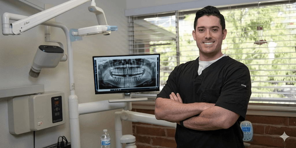
Our cone beam computed tomography (CBCT) unit takes dental imaging to an entirely different level. Where traditional two-dimensional images show us a flat representation of complex three-dimensional anatomy, a CBCT scan produces a detailed, three-dimensional image of the teeth, bone, airways, and surrounding structures — revealing detail that simply cannot be seen any other way. The radiation dose of a CBCT scan is comparable to that of a traditional panoramic film image — making it a powerful diagnostic tool that does not come at a significant cost in exposure. Despite this, we use CBCT imaging selectively and purposefully, reserving it for cases where the additional information will meaningfully impact diagnosis or treatment planning. In our practice, CBCT imaging plays a critical role in implant planning, TMJ evaluation, airway assessment, complex restorative planning, and the early detection of pathology — including developing abscesses that may not yet be causing any pain or symptoms. The ability to identify these issues before they become emergencies is one of the most valuable aspects of this technology, and one that patients often don’t expect. When a patient needs to be referred to a specialist, their CBCT images go with them — providing the receiving provider with a comprehensive, three-dimensional view of the relevant anatomy from day one, improving the quality of specialist care and eliminating the need for duplicate imaging.
Two-dimensional X-rays and three-dimensional CBCT imaging each have their role, and we use both thoughtfully. What they share is this: they give us the information we need to provide care that is accurate, safe, and truly in your best interest. In our practice, proper imaging is not negotiable — because proper care isn’t possible without it.